Anatomy And Physiology Of Brain Pdf
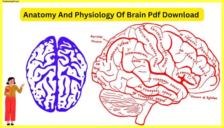
Today in this article we will discuss the Anatomy And Physiology Of Brain Pdf with examples and definitions, we will try to clear all of your doubts about Brain Anatomy And Physiology in this article So Let’s Start.
The Brain
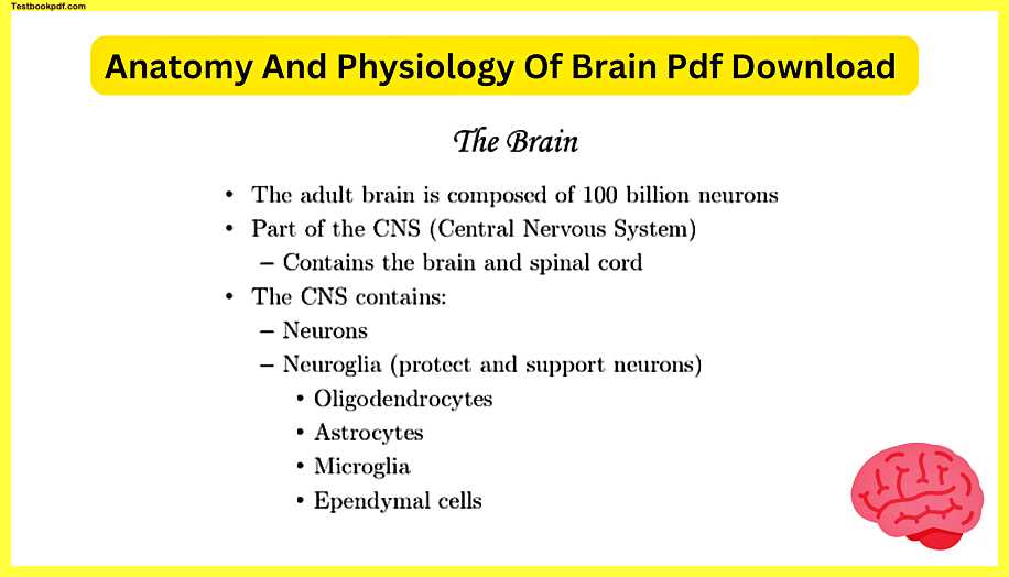
The adult brain is composed of a hundred billion neurons which are amazing to think about that’s a lot of cells in the first few years of life you actually lose a lot of brain cells think of it this way.
A baby their brain is like a giant untamed bush and you don’t need all of those little leaves and all those little branches to have the bush you know look nice and serve a purpose.
So imagine that the baby brain undergoes a lot of pruning you kind of clip certain parts off that aren’t necessarily needed and it becomes this nice functional shape with compartmentalized parts so actually proportionally baby brains have a lot more neurons than they need and by the time you get into your adult life you’re not really making hardly any neurons anymore the ones you have are the ones you’re gonna use until the end of your life.
The brain is a part of the central nervous system and we’re gonna breathe II that in the future as the CNS and the central nervous system unlike the peripheral nervous system contains the brain and spinal cord it’s literally the central brain and spinal cord going down your back in the middle in addition to just your neurons that make up a lot of your brain you’re also gonna have what are called neuroglia and these are specialized kinds of neurons that don’t do just the regular signaling that they don’t just do the sensory and motor part of how the CNS works with your body the first one is Oligodendrocytes.
Oligodendrocytes
Oligodendrocytes came up in the previous set of lessons regarding how neurons work so if remember Schwann cells being those myelin sheaths wrapping around neurons in your peripheral nervous system that branch off from your spinal cord and brain oligo Dentro sites they make those wrappings inside of the central nervous system so they help insulate and make saltatory conduction possible in the brain.
Astrocytes
They maintain the blood-brain barrier they have a lot of functions but I find this the most intriguing that astrocytes which kind of look like little stars that’s why they’re called astrocytes looks like there’s like little kind of beams extending from their little cell body astrocytes maintain the blood-brain barrier and interesting lis enough your bloodstream not everything in there is able to go into your brain it’s a protective mechanism which is very important and actually some drugs introduced into the bloodstream they stay out of the brain and they affect other organs other nerves outside of the central nervous system and then how things can pass through one of the many other functions of your blood-brain barrier is protecting your brain from certain microbes certain parasitic worms and interestingly enough there is actually a kind of parasitic worm I’ve heard of that has evolved the ability to dissolve the blood-brain barrier so it actually does end up inside a person’s brain if it gets into their body.
Microglia
Microglia is another kind of neuroglia think of microglia as the trash men then the trash compactors of the brain they’re kind of like a macrophage you’d in your bloodstream an eater of cell debris so they go around doing what’s called phagocytosis where they grab stuff outside of the cell outside of themselves like let’s say it’s waste that’s accumulated it might be some kind of foreign invader that got in there and it shouldn’t be there so they’re cleaning up the trash in your central nervous system.
ependymal cells
You’re gonna find these more often around the ventricles which are the little hollow cavities you’ll see more about those in the future on this presentation they’re like little holes little cavities inside the brain and the central canal another cavity that’s gonna find in the spinal cord you would find these cells adjacent to those spaces deep within the central nervous system because they help produce what’s called cerebrospinal fluid and they help regulate it and kind monitor it in case something goes wrong and they and they do help produce more of it if it’s needed or produces less of it if there’s already plenty.
Brain Development
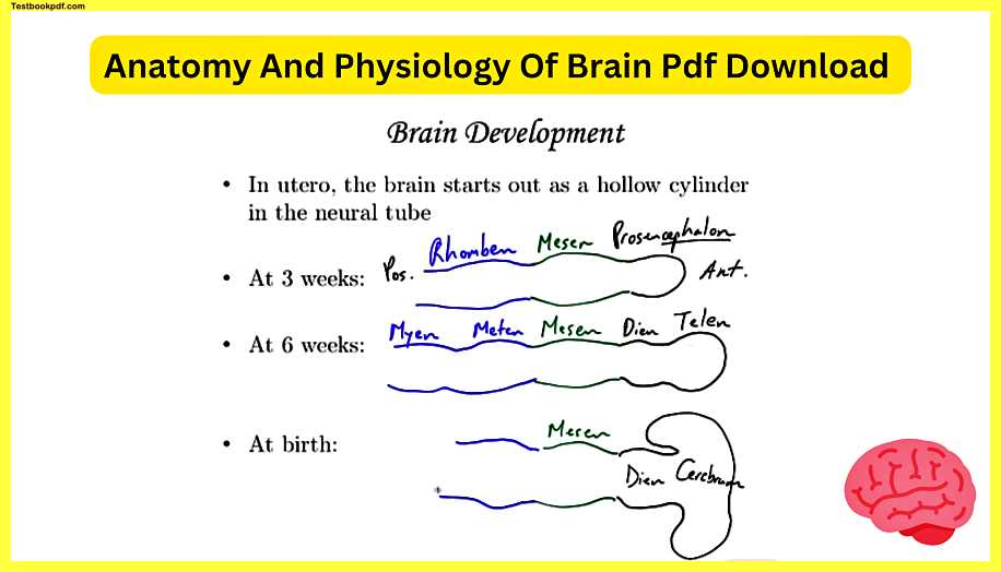
When it comes to brain development you start small if you look inside a tiny little embryo on a microscopic level you have what’s called a neural tube that first develops and it’s just very simple sets of neurons that are lined up and there are a hollow section in the middle so it is like a tube that tube ends up getting little pockets you can call them brain vesicles if you want.
At 3 weeks
So at three weeks let me actually color code this for you you would see three main sections and up here this is the anterior part towards the front and back here is going to be the posterior part up at the anterior part you would call this first bump the pros and Cephalon pros and Cephalon and this word Cephalon is going to be the suffix the ending of all of these little areas so pros n Pro meaning before like prologue is up at the front the next part I’m going to do in green is actually gonna be called the mesencephalon and just for the sake of time and making.
It’s simpler I’m just gonna write mezzotint it has Cephalon after it of course and then finally at the back end the posterior part this is the Raman Cephalon and that’s at three weeks usually before a woman even know she’s pregnant this has already happened pros and Cephalon mesencephalon Robbins Cephalon.
At 6 weeks
At six weeks it gets a little bit more developed and expanded so I’m gonna still use black for what happens to this anterior vesicle it actually becomes two areas it grows into called the diencephalon and telencephalon.
Next up you’re gonna see the telencephalon actually gets to be more like a tea it actually will expand on the sides here and if you use your imagination kind of take her to the side if this expands horizontally you can see how it looks like the top of a tea that’s how I remember it the middle part called the mesencephalon.
Just stays the mesencephalon and actually the mesencephalon is a part of the adult brain so this is not gonna take on a different name as we go to the parts of this brain tube if you want to call it that up until birth the ramen Cephalon does become a couple of other areas and there’s a trick to remembering these areas this is the mech ten Cephalon and this is the Mayan Cephalon the way that I remember the back end of this is its alphabetical mess in with an s met n t comes after s and then Mayan so s t and then doesn’t even go to eat it goes my so these are in alphabetical order that’s how I remember them from the middle part to the posterior portion and then we’re gonna jump ahead way further than
At six weeks we’re gonna jump to 40 weeks which is the approximate time that it takes for a baby to develop and then be born you’re gonna see some names that you probably have seen for having to do with brain anatomy.
At Birth
So the front gets a lot bigger I’m not even doing it just as how big this gets the telencephalon becomes the entire cerebrum the cerebrum in an adult brain or a newborn baby brain is that recognizable part with all the wrinkles on the surface that’s a lot of brain tissue so this becomes what’s called the cerebrum this is still the diencephalon and later on in this presentation you’ll see the diencephalon is just deep to what’s going on in the cerebrum the mesencephalon stays the mess and Cephalon that’s like an area between the diencephalon and the brainstem so this is still the messin the Med encephalon becomes two parts that are kind of in the inferior / posterior parts of the brain this becomes the pons you’ll hear about that more later and the cerebellum cerebellum actually means mini or little brain in Latin because it’s kind of a modified version of the word cerebrum and then last we’re still using blue this last part here becomes the medulla oblongata which is a major portion of the brainstem leading to your spinal cord so by the time a baby is born a lot has come of this initial simple neural tube these brain vesicles become well-established and enhanced as time goes on.
Superficial Brain Structre
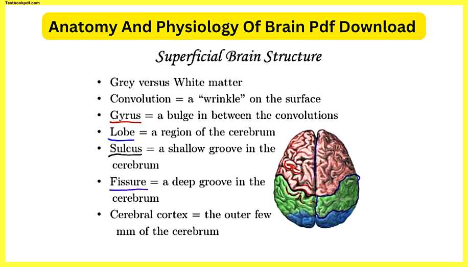
When we look at the superficial brain structure in an adult or a young kid for that matter on the superficial part what you’re looking at here we’re actually looking at a top view superior view down on the cerebrum here’s the left hemisphere the right hemisphere you’re not gonna actually see pink green and blue this is color coded so you can see the different lobes which we’re gonna cover in a little bit but on the outside they nicknamed it gray matter it’s darker if you cut into the brain and look deeper you’re gonna see a greater proportion of white matter the gray matter tends to be more of the parts of neurons that are cell bodies and what makes it great is all the cell bodies compacted together the nuclei which are slightly darker all of those together give it a slightly darker appearance on the other hand the white matter that tends to be deeper in the connecting parts that get the parts of the brain together and signaling each other that’s more white and the white comes from more axons remember the axons are those long parts that are the signaling parts for neurons to communicate with another neuron or another part of the body so that white comes from the myelin sheaths and in this case you’re gonna see a lot of those oligodendrocytes covering those parts of the axons and that has more of a white appearance.
Convolution
Convolutions are the technical terms for the wrinkles people say you know every time you learn something you get a new wrinkle in your brain that that’s an oversimplification and kind of a cartoony way to put it it’s not like oh I just learned something new and then a wrinkle just magically appears at that moment there is some truth to it because the more wrinkles you have the more neural tissue you’re fitting within your skull to think about the surface of this having no wrinkles that just means way less surface area so you can compact a lot more oral tissue by folding it up and having it folded in this in this very nice way so yes as you do as your brain grows and as you have neurons being made and then other neurons dying off because they’re not needed they’re not as important you’re going to have wrinkles established and maintained a gyrus.
Gyrus
Gyrus is basically a Ridge or a bulge so the gyrus we’re gonna color code that red here’s a gyrus right here that’s a bulge here’s another bulge this gyrus here that extends in this red part that actually has a very specific name it’s called the precentral gyrus because this black line here is called the central sulcus so because it’s in the front of it they actually call this the precentral gyrus, on the other hand, this right here this gyrus is called the postcentral gyrus because it’s just behind the central sulcus plural for Gyrus.
lobe
A lobe is probably a more familiar term all of this green area is a lobe and this is specifically the parietal lobe the red ones are the frontal lobes the blue ones are the occipital lobes you can’t see it in this image but the temporal lobes are below like I mentioned earlier a sulcus I’m gonna do.
Sulcus
This is black a sulcus is a Deep Wrinkle it’s a very obvious Ridge or sorry I’m not Ridge an obvious kind of sinking in if the ridges are the gyri in between them you can call that a sulcus so it’s a shallow groove it’s not very very deep that’s a fissure.
Fissure
So a fissure will do that one in purple is like a sulcus but way deeper like a ditch here is one of the biggest fissures in the brain this is called the longitudinal fissure and that’s the obvious barrier that separates the two sides of the cerebrum
Cerebral cortex
The cerebral cortex you’re looking at it the outer few millimeters of grey matter that’s surrounding the cerebrum is called the cerebral cortex you have a lot of neurons there a lot of mental processing and that’s where all of those human things we recognize on a daily basis come from you have a lot of higher processes going on there at the cerebral cortex level so we look at the cerebrum as a whole it is the higher brain it’s the most obvious part of brain anatomy when you look at it this image you’re seeing here is actually an fMRI functional magnetic resonance imaging picture of my brain so we’re looking at my brain this is a sagittal section.
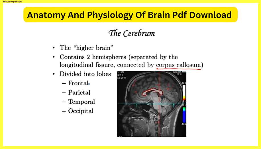
So it’s as if like we took a slice right through here and you could see my profile there here is the cerebrum all of that and here is the deeper parts we mentioned diencephalon earlier this is the diencephalon right in there here’s the pons medulla cerebellum but all of this the majority of the brain is the cerebrum and actually this image it’s like you cut straight through that longitudinal fissure the corpus callosum which you’re gonna hear more about later is this white strip and that’s hundreds of millions of neurons specifically axons because it’s white that are connecting the two sides of the cerebrum so the way that the left and right hemispheres communicate is through this little white strip and the cerebrum is divided into lobes as I mentioned earlier the frontal lobe up front so it’s a large lobe parietal kind of off to the side up top temporal lobes you can’t see them in this image they’re on either side here if you were seeing the actual left side of the intact brain or the right side they’d be visible and the occipital lobe tucked back here.
Frontal lobe
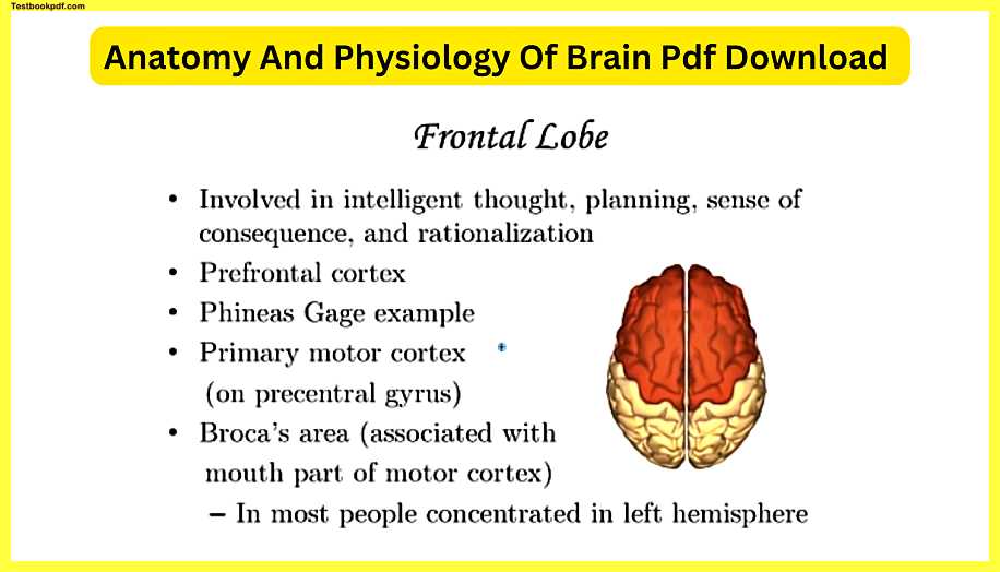
So the frontal lobe this is a superior view you could see they’re they’re quite significant you’ve got the left side and the right side of the frontal lobe the frontal lobes are involved in intelligent thought planning sense of consequence and rationalization these are the parts of the brain that really make you intelligent so if you’re thinking and you’re tapping here you’re you’re tap in the part that’s gonna help you either solve the problem or come up with an intelligent answer the prefrontal cortex is actually the very very front part of this frontal lobe and that’s where most of that intelligent stuff is going on the example of Phineas Gage is is a really good way to think about what the frontal lobe does Phineas Gage was a railway worker back in the 1800s and there was this freak accident that happened when they were laying down track they had an explosion that had to do with like getting stuff deep in the ground to hold the rail down and there was a misfire and hot rod just went through his head it actually didn’t hit any major arteries it missed one of what would have made him blind and missed a lot of areas but it went straight through the frontal lobe and out the top of his head and if you’re wondering why didn’t he bleed to death the heat from that thing that rocketed through his head just Carter eyes just kind of sealed shut blood vessels with all that heat.
I’m sure he was in some kind of hospital for a while but he lived and this was the first time in recorded medical history that we know of in which somebody had just damaged their frontal lobe and that’s it so Phineas before the accident was a polite gentleman he showed up on time to work he had a great personality people liked him and after the accident people commented that Phineas was not Phineas anymore he tended to curse a lot more he tended to show up late for work those things that made him who he was in terms of recognizing a responsibility having intellectual responses caring how his actions impacted other people that was gone so think about the frontal lobe as something that that made Phineas who he was and once that accident happened he acted more like kind of an uncivilized person the primary motor cortex is tucked back in the posterior section so the motor cortex here and here that actually controls the muscles of your body so along this Ridge you actually have sections that are devoted to moving the arms moving the fingers moving your legs moving your torso your back and really the initiation of all of those movements comes from this Ridge in this Ridge and remember it’s called precentral gyrus because it’s in front of that central sulcus and the amazing thing is the amount of tissue that’s devoted to your fingers and your mouth is fortunately larger than you would expect you know your legs and your back are large proportionately in your body but the actual sections of this primary motor cortex the initiation of the motor movements of the back and the legs is is significantly smaller than your fingers and your mouth but think about the really precise movements you need to do with your fingers or the precise movements you need to do with your lips your tongue in your jaw to speak and all the things to do with your hands there’s a lot going on.
So the amazing thing is they’ve actually made these little neural maps showing how proportionately these parts of the motor cortex control the muscles and all the parts of your body Broca’s area is an area associated with the part of the motor cortex that controls your mouth but the reason why it’s a little bit different is Broca’s area has to do with coordinating those mouth movements in a way that enables you to speak clearly so somebody with damage to Broca’s area can still make noise they can still say words but it’s not in a coordinated way and of course it varies depending on how badly it is damaged but some people with a problem with the Broca’s area they just they can’t find the right words or they can’t speak words in a clear way that enables other people to understand them and in most people this Broca’s area is concentrated in the left hemisphere rather than in the right and we’re gonna come back to that later about how that does impact certain situations.
Parietal lobes
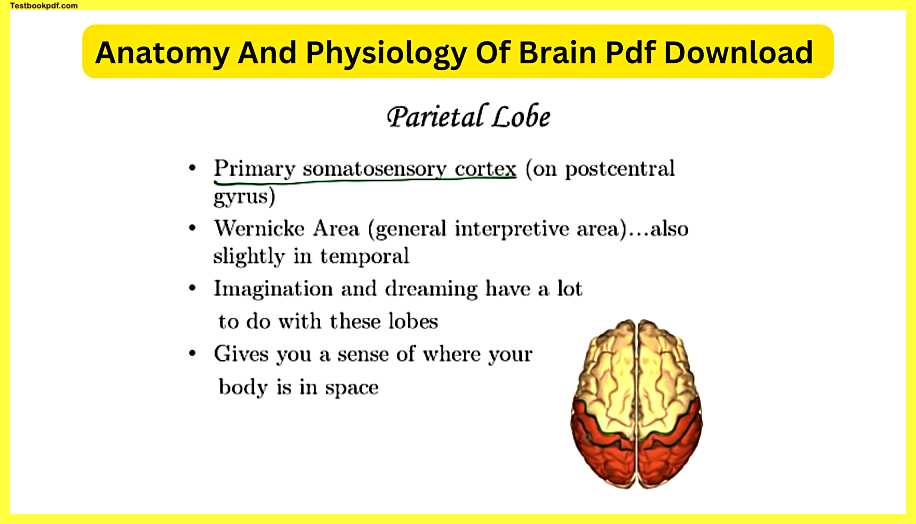
The parietal lobes conversely unlike the frontal lobe have a strip that’s for sensing so remember this where I’m tracing over right now that’s the precentral gyrus which allows you to coordinate muscle movements but just on the other side of the central sulcus you have the primary somatic cortex or somatosensory cortex that’s right here and typically if if in this part here you have the ability to move parts of your arm just on the other side of that sulcus you have the parts that allow you to feel someone actually touching or something touching your arm so all of the feelings you get something touching your lip something touching your head something touching your back whatever part of your body is being touched even stuff inside your body that you have a sense of those signals are coming up into these parts of the parietal lobe and just like with the motor strip or the motor cortex the proportion of these strips that actually has to do with sensing stuff on your hands and sensing stuff on your lips those stimuli are much more obvious which makes sense as humans we do a lot of touching with our fingers and we want to be able to tell one object that’s different from another and in a fine-tuned way.
So the proportions yeah for the hands and for the lips greater than the actual size of them on our body and then there’s there’s been these interesting drawings made where they draw what would a person look like if we took the somatosensory cortex and took the proportions in there and then drew that that individual those drawings they have a large face huge lips the hands are humongous the feet are kind of big too and the torso the arms and the legs are all kind of puny but you see this person with large hands and large lips because those areas are the most sensitive and while we’re on the subject of hemispheres it is true that the left side of the body communicates with or sorry I just said it backwards the left side of the cerebrum communicates with the right side of the body and vice versa the right cerebrum that hemisphere communicates with the left side of the body wernicki area or Wernicke’s area is an interpretive area that’s mostly in the parietal lobe but it’s also slightly in the the temporal lobe as well if you want to what this area does it has to do with the temporal lobe a bit because the things you hear those sound waves go into your ear and the nerves connect what you hear to the brain in the temporal lobe and then that’s how you actually oh I can hear it there’s an area of the temporal lobe communicating with this part of the parietal lobe and it’s called Wernicke’s area named after the doctor who discovered it Virna key would probably be a better pronunciation but this area allows you to hear speech as something that you can understand.
So if you have damage to Wernicke’s area something that could come as a result is you may still understand what the word sit means you may understand what the word here means but if somebody said sit here sit here the person with damage to Verna Keys area would be like they they wouldn’t understand really what that meant when those two words together so the ability for us to understand what words actually mean when they’re strung together that’s thanks to Verna Keyes area imagination and dreaming have a lot to do with these lobes I’ve read that when they analyze einstein’s brain his parietal lobes were actually larger than the average person’s brain so maybe he had a genetic gift more so than others in terms of understanding or visualizing how the universe works and the parietal lobe gives you a sense of where your body is in space which makes sense because if that somatosensory cortex or somatosensory strip is telling me what parts of my body are being stimulated having my feet touching the ground and having my arms touching this table that I’m at that has a lot to do with the parietal lobe just my body realizing where my arms are where my feet are if I do a headstand that’s gonna obviously a very different set of signals going up into my brain.
Temporal lobe
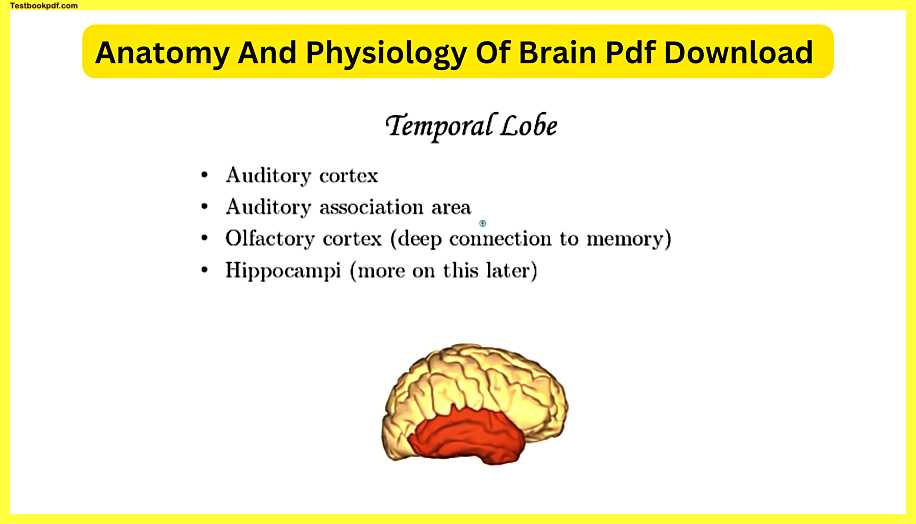
The temporal lobe is mainly for hearing but that’s not all so that’s why you see the auditory cortex here and here’s the right temporal lobe on the opposite side you’re gonna see a very similar lobe that’s the left temporal lobe so the auditory cortex that’s where the nerves from your ears go to and this is the one part of the brain where it’s not switched so with my left hand feeling something via touch those signals are going up to my right part of the cerebrum the right hemisphere but when I hear something through my right ear that actually just it doesn’t go to the other side it just goes straight to the right temporal lobe and same with the left ear the auditory Association area allows you to make sense of what you’re hearing.
So that is communicating with Verna Keyes area also words have a meaning to them beyond just what the definition is there’s, of course, the connotation of what a word means you can say things in very different ways and if you had damage to the auditory association area that inflection that people apply to different words may not make entirely it may not make sense to you the olfactory cortex is the parts of the temporal lobe associated with smell.
So when we talk more about smell in future lessons you’re gonna see that the olfactory tracts from the actual receptors that allow you to smell go right through the temporal lobe and they pass right by the hippocampus and pleura would be hippocampi people camp I have a lot to do with memory and you may have heard that if all the senses smell has the deepest connection to memory because there is truth to that the fact that the olfactory cortex goes right next to these parts of the brain that allow you to have those long-term memories yeah that’s why you know if you smell freshly baked cookies you may immediately think about your grandmother’s cookies that she made years ago and it just triggers that response you may smell a perfumer cologne that reminds you of somebody immediately and so that’s some examples of how smells could trigger long-term memories.
Occipital lobes
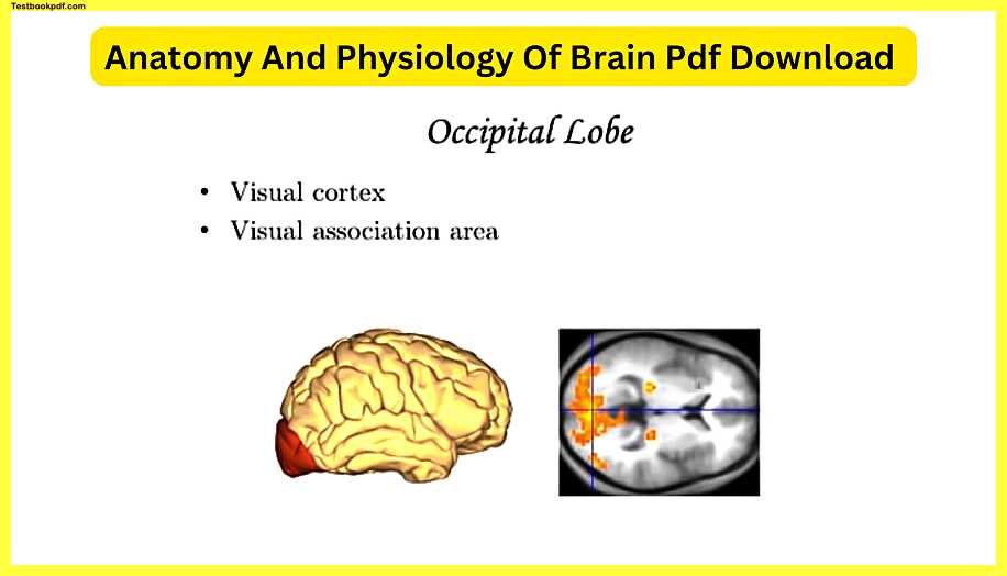
The occipital lobes mainly for vision the visual cortex is the part of the the back occipital lobe of the most posterior part of the cerebrum that actually just enables you to see the visual Association area is what’s allowing you to make sense of what you are seeing you know so if you have damage to the visual Association area you may still be able to see perfectly fine but when I look at a sentence and I see let’s let’s see I see the other word B e di seabed III CBE D and since I don’t have a problem with a visual association area I know that means bed but someone with damage to this the association area they’ll be able to see the B see the E and see the D but not make sense of what those all mean together which would be a terrible thing back here this is actually one of those horizontal sections or a transverse transverse cross section through the cerebrum all of this yellow stuff is the parts of the occipital lobe that are lighting up that are actually functioning and using sugars and and doing all those action potentials to enable a person to see so they’ve done these time and time again showing how these parts of the brain are associated with seeing and then interpreting what you’re seeing.
Corpus callosum
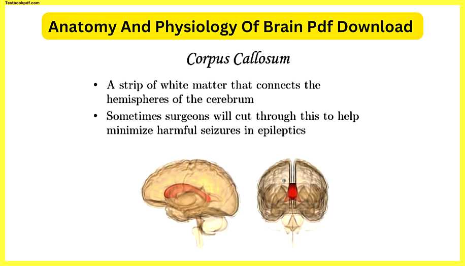
The corpus callosum is a strip of white matter like I said before that connects the two parts of the cerebrum here’s a nice side view of what the corpus callosum is and here’s a frontal view if you could look through the frontal lobes and see it you can see that here’s that longitudinal fissure that that deep kind of ditch between the two sides of the cerebrum. the two hemispheres but yeah you have a connection between the two with a lot of axons hundreds of millions of axons that are allowing the left and right side to communicate sometimes through surgeries the doctors will actually cut through it one of the reasons.
Epilepsy seizures
Why somebody who has epilepsy seizures? who seizures that come again and again and again seizure is like kind of an out control electrical storm in a person’s brain and seizures over the long run can do a lot of harm so they’ll cut through that because somebody who gets a little you know seizure starting in a part of the brain on let’s say the left side it will jump to the other side through the corpus callosum so if you cut off the connection it prevents the seizure from jumping back and forth and creating some harmful scenarios some people who are epileptic if they cut through that you get some interesting effects because.
Now it’s not communicating as well you don’t have the left and right hemispheres kind of communicating them here’s an example if somebody who has a cut corpus callosum is holding a pen in their left hand remember the left hand it’s communications with the right hemisphere now the parts of the brain that have to do with naming objects like you know making sense of words is more on the left side of your brain.
Now that that has to do with this right the left side of the brain has to do with my right hand so a person who holds this in their left hand with a cut corpus callosum they’ll be able to know that they’re holding this describe the object but they’re probably gonna have trouble naming it because the left-hand doesn’t have the signals going up to that language part of the brain on the left side conversely if they then take that object and put it in the person’s hand on the right side well.
Now, this goes up to the left part that your language area tends to be more on the left side so they’ll say yeah it’s a pen but then if you ask them is this the same object you were holding in your left hand the person probably won’t be able to tell you yes or no so it’s an interesting result from cutting the connection between the left and right sides of the cerebrum and actually there’s an interesting a side note here there’s one other area in the very front called the anterior commissure and that commissure connects a little bit of the front parts of the cerebrum so people who have a split disclose them you still have one little area in front of it where you can have some communication so the seriousness of what I was just talking about can vary from person to person.
Limbic system
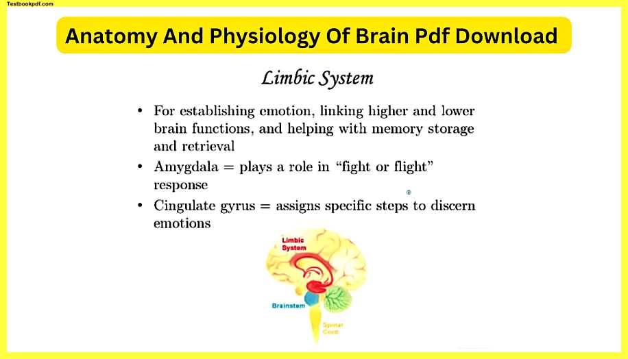
The limbic system is deep within the cerebrum and it’s actually on both sides I thought it’s slightly on the left slightly on the right it’s for establishing emotion linking higher and lower brain functions and helping with memory storage and retrieval the amygdala is a major part of it that tends to be in the deeper section this whole red stuff colored in that’s the limbic system so the amygdala plays a role in the fight-or-flight response which you’re gonna hear a lot more about in future lessons that I’m gonna give you if you had a damaged amygdala it’s gonna be hard for that person or impossible to connect emotion with the response for instance if I step out into the street and out of the core of my eye I see a car coming that’s going to initiate a very quick response in my body to back away somebody with a damaged amygdala might have some trouble with that happening recognizing that something is threatening to you is thanks to the amygdala and that’s really when the fight-or-flight response is gonna come into play something is threatening to you and the amygdala is connecting your response with the higher parts of your brain that gets you to react effectively and keep yourself alive the cingulate gyrus or the cingulate cortex is on the top part here where I’m tracing over and this part of limbic system assigns specific steps to discern emotions.
So if you showed somebody a bunch of pictures of faces and they have a problem with their cingulate gyrus you could show the person a picture of a face that’s like this so you could tell since presumably, you don’t have trouble with out-party your brain that I’m giving kind of a surprise or scared look the person who has damaged they’re looking at that part may not be able to tell you that they’re scared or surprised they may look at the open mouth and say um I think they’re hungry because they’re missing that part that that’s enabling them to look at a face and figure out what emotions that person is experiencing and that’s part of being a social being as humans we rely a lot on what other people are feeling or experiencing to inform us of what’s going on in the environment so the limbic system is very important for keeping human beings alive.
Hippocampus
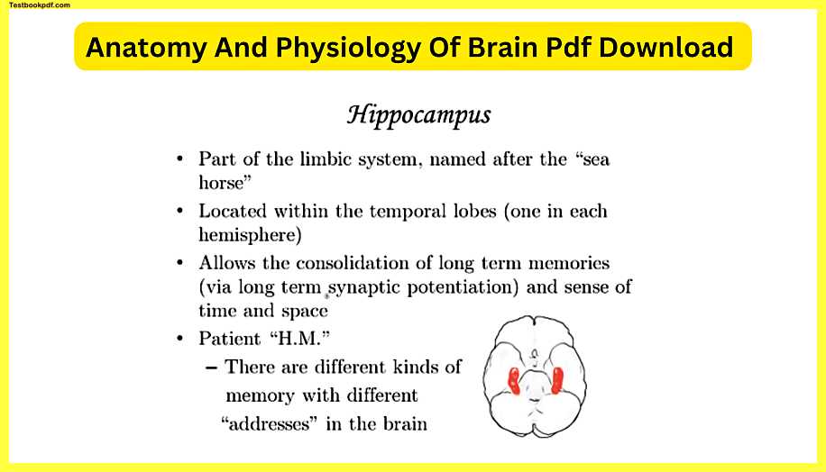
The hippocampus also called hippocampi if you look at the plural word is a part of the limbic system in in the lower areas and it’s like two little kind of nodules two little areas deep within the temporal lobes named after the seahorse so hippocampus literally means seahorse I guess if you use your imagination maybe you might see that but here are the hippocampus and we’re actually looking at at a inferior view of the brain here so here is temporal lobe temporal lobe you can see that they’re concentrated in those sections the hippocampus has to do with getting memories and allows the consolidation of long-term memories via long-term synaptic potentiation and a sense of time and space let me give you some examples so long-term synaptic potentiation that’s a fancy term for saying that you’re gonna get a set of neurons in a connected pathway staying put so that this one has the ability to stimulate this one permanently if need be so a long-term memory is because you have a neural pathway established in your brain that there’s some importance to it might have emotional importance I have importance for just remembering your phone number remembering your address so those things that you remember for years you can thank the hippocampus because the hippocampus has enabled through connecting to them enabled those parts of your brain to maintain neural pathways.
Now short-term memories they form and they go they form and then go for instance if you asked me what I had for lunch on Tuesday of last week I don’t remember I could guess but chances are I’d be wrong there wasn’t a need for me to remember what I had for lunch on Tuesday last week now some people have a gift have something special where they can remember every day of their life and they call that a disorder it is somewhat of a gift but the average person you’re only gonna be having long term memories maintained when they’re needed so a short-term memory if you have a neural network established for let’s say a conversation you had that wasn’t of huge importance with someone after a day or two that conversation the minor details of it you won’t need that network anymore it could be reestablished for some other new experience that you just had so the brain has a certain amount of what’s called plasticity this is an important word that will come up later plasticity meaning the way that the brain is arranged is not set in stone you can actually adjust what it’s doing as your life changes over time and that’s very important prior science from years ago 30 40 years ago they really thought that the adult brain was kind of set in stone and research lately has showed that’s not true the brain has a great degree of what’s called plasticity patient HM is the best example I could give you for how the hippo can’t by work and and when he was alive for confidentiality reasons he was just called HM but his his name.
We now know is Henry mOLAISON and Henry mOLAISON has passed away so that’s why we know more about him now he is one of the most studied patients in medical history years ago decades ago he had his hippocampus surgically removed in operation because of issues with something about his brain but the importance is that every other part of his brain was functioning properly but he’s missing the hippocampus so that’s a great way to study what this does I mean that’s how we figure out what parts of the brain do if you damage one part of it but the rest of its intact you can figure out what’s missing or what’s different so inpatient HM and Henry with the doctors that studied him for years he wouldn’t remember them they would come in weekly and study him and he’d have to you know be reintroduced to these people he could remember everything up to the surgery so you could ask him about childhood memories things about his family he would remember those completely but once the hippocampus were removed once the no longer functional that prevented him from making long-term memories that we take for granted the interesting thing is that memories have different addresses in the brain.
So I don’t want you to think he was completely incapable of long-term memories it depends the amazing thing is that with the house that he lived in after the surgery he was still being studied while he was living there with a caretaker if you asked him to design the floor plan he can design it exactly if you ask him to do it and theory behind that is even after his hippocampus was removed he had walked around that house for years every day same house so something else in his brain was allowing him to remember that another interesting thing about that is I I don’t know if this is a tall tale I heard this story about HM that the doctors went in one day and shook his hand you know let’s say was four doctors they all shook his hand and introduced themselves as if he had never seen them before a few days later the doctors come back in shake his hand and now one of them has one of those little buzzers from those shocking buzzers on their hand so the fourth doctor shakes his hand introduces himself and shocks him and he of course is disturbed by that and maybe they all had a good laugh right afterwards so you would think that he wouldn’t remember that at all the next time those four doctors come in the last doctor that walks into the room still has the buzzer on his hand so he he meets the first three doctors meets first three the one comes and hm kind of hesitated a little bit in meeting this person so maybe some other part of his limbic system had allowed him to retain some kind of emotional response to individual and then just this just goes to show you that the brain is very complicated even if you’re missing the hippocampus it doesn’t mean all of your long-term memories are not gonna happen anymore but the majority of it is gonna be affected.
Basal nuclei
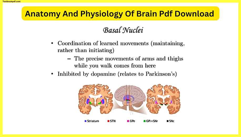
The basal nuclei has to do the coordination of learned movements and it’s it’s maintaining those learned movements over time rather than initiating it so for instance when you walk the precise movements of your arms and thighs you don’t really have to think about it consciously when you walk your arms just kind of sway back and forth opposite of your legs right when your left arm moves forward your right leg tends to move forward and so on and so forth we don’t even put conscious thought into it but you can thank the basal nuclei these these regions deep within the brain and you can see they’re concentrated in the white matter the signaling parts which makes sense because they’re signaling these coordinated movements of your arms and legs while you’re walking parts of the basal nuclei are inhibited by dopamine I brought this up earlier in a previous lesson that if dopamine is the neurotransmitter that’s turning off some of these parts if you don’t have as much dopamine being released as you should you could get some uncoordinated movements making it hard to make precise motions.
Olfactory bulb/tracks
The olfactory bulb/tracks have to do with smelling and this is a great image from Gray’s Anatomy we’re looking at an inferior diagram of the brain these little red parts outlined there look like little white strands they’re very obvious when you, when you turn a brain underneath you, can see these two parts and they have to do with receiving smells from what is called the olfactory receptor cells and bringing those parts into the brain these are the only nerves that go directly into the cerebrum rather than the diancie on our brainstem so if you think about the nerves coming from your arms from your legs from your eyes they go through kind of like relay stations on the way up to your higher brain the exception is these olfactory tracts and they lie just inferior to the prefrontal cortex like I mentioned before the very front part of the frontal lobe is called the prefrontal cortex.
Ventricles
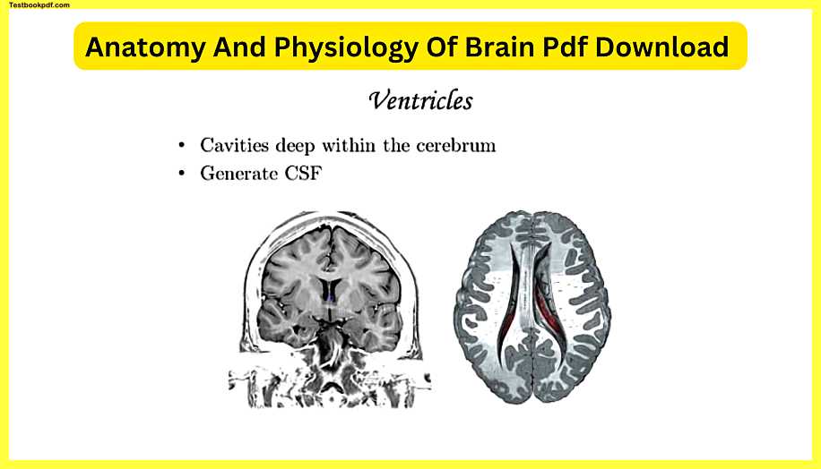
The ventricles of the brain are basically cavities if you’ve heard of the ventricles of the heart the the hollow areas within the heart it’s the same kind of structure in terms of why they’re named this these are hollow cavities deep within the brain so we wouldn’t call this a ventricle up here remember this is the longitudinal fissure that you can see very easily when you look down on the brain from a superior view this is a ventricle and this is a ventricle so this is one of those frontal or coronal cross-sections straight through the cerebrum and this gives us a good view you can see part of the ventricles here this is a transverse or horizontal section so if you wonder what do these cavities do they have to do with generating and circulating the CSF also called the cerebrospinal fluid and there’s actually a kind of cell known as it was actually a region known as the choroid plexus that’s adjacent to ventricles and remember the ependymal cells I almost forgot the word the ependymal cells they have to do with making the CSF and regulating it so these ventricles those cavities within the brain get CSF in there and then they circulate that around the outside of the brain and the outside of the spinal cord as well as inside was called the central canal which will come up in a later lesson but what is this CSF do what is it why is it important well it has a lot to do with protecting the central nervous system and helping to regulate the amount of gases coming in and out the wastes leaving nutrients coming in so that the health of the CSF impacts circulation in and out of the brain very important.
Diencephalon
The diencephalon is deeper than cerebrum so now we are done with the cerebrum that was a lot of slides but the cerebrum is very complicated so if you go just deep to that there are two regions the thalamus and the hypothalamus hypo literally meaning below the thalamus and the thalamus is kind of like a relay station it brings sensory information up into the cerebral cortex and in a coordinated fashion and like I said earlier the majority of the nerves coming into the central nervous system they have to go through the thalamus on their way up a classic example that is the optic nerves the hypothalamus which is directly inferior to the thalamus has a lot of roles it’s just above the pituitary gland and you’re gonna see this more obviously in future lessons if you take a cross-section of the brain there’s a little thing that looks like a pea that’s hanging from the base that’s the pituitary and the hypothalamus is what is right above it feeding reflexes salivating a baby making certain motions during breastfeeding has to do with the hypothalamus initiating those things the feeding Center and thirst center you getting hungry and thirsty the signals from the brain that have to do with that come from the hypothalamus circadian rhythms have to do with sleep versus being awake so typically for most humans when the sun goes down after that happens you tend to get more tired the exception being if you work a night shift but for the average human being circadian rhythms has to do with signals from the environment telling your brain it’s it’s sleepy time and then when the Sun comes up it’s time to wake up.
So the regulation of the circadian rhythms that have to do with you know changing your conscious state that’s from the hypothalamus communication with a pituitary gland if you remember me saying that the pituitary gland is like a little peaked gland hanging right below the hypothalamus a lot of people refer to the hypothalamus is like the master of the endocrine gland it’s like the big head honcho and the pituitary gland is kind of like its second-in-command the hypothalamus tells the pituitary gland to secrete certain hormones at certain times and the pituitary gland impacts the other endocrine glands causes them to release hormones or the pituitary gland just causes other organs to be regulated for instance of the pituitary gland secretes growth hormone.
So making your bones elongate in the proper way throughout your adolescence that’s regulated via the hypothalamus and pituitary gland and subconscious skeletal muscle control all those movements you do that you don’t have to think about you can thank the hypothalamus the pineal gland is the final part of the diencephalon it’s actually in the posterior portion of this area it’s like a smaller pea it’s actually a similar looking structure but it’s not hanging from the brain it’s tucked in the back this has to do with secreting melatonin and melatonin is a neurotransmitter that actually puts you to sleep and there’s been a lot of studies done with people taking melatonin you know pills when they go on a plane to make them go to sleep and I read a study that actually had to do with the placebo effect of that suggesting that the average person who takes a melatonin supplement didn’t meet it that the melatonin in your brain is actually enough to put you to sleep naturally.
Mesencephalon
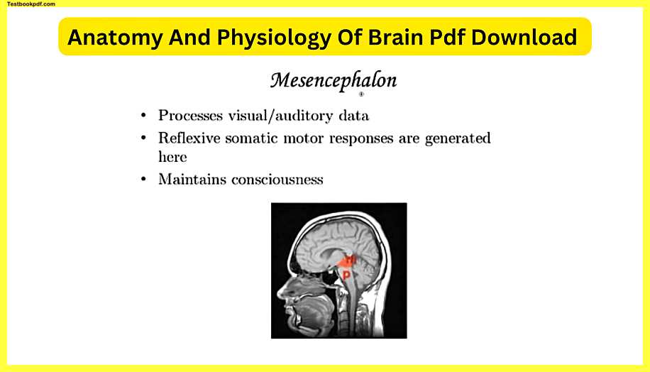
The mesencephalon is just kind of posterior/inferior to the diencephalon right here in this cross-section this is actually not my head anymore but this is another sagittal cross-section straight through the corpus callosum and you can see this M is that mesencephalon has a lot to process visual and auditory data that comes in from the eyes and ears your reflexive somatic motor responses are generated here and that’s fancy terminology for when you hear a sound or see something on the corner your eye and you react so if I heard something over there and I did this without even having to think about it that’s because of the mesencephalon so if you if your eyeballs move to an in the reaction of something has a lot to do with the mesencephalon and the maintaining of consciousness comes from this area so I’m using my mesencephalon right.
Pons
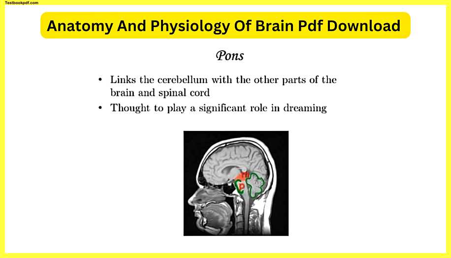
Now next to the pons same image as before the pons is this bold right here that’s the pons so it’s just below or inferior to the mesencephalon and just anterior or in front of the rest of the brainstem so this links the cerebellum which is right back here with the other parts of the brain and spinal cord and considering its proximity to where it makes sense that it would do that so the pons has a lot to do with getting communication between the higher brain and lower parts of the brain and it’s thought to play a significant role in dreaming.
Medulla oblongata
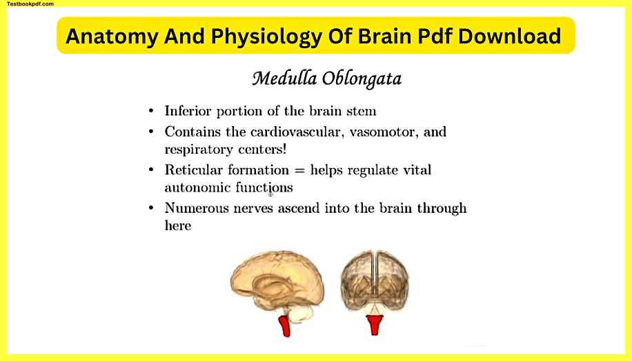
The medulla oblongata is the majority of the brainstem so you can see that it is the inferior portion just below the the pons so here’s a side view here is a frontal view or anterior view it contains very important here the cardiovascular vasomotor and respiratory centers damage to the medulla oblongata often results in a fatality because your heart rate is controlled by the Cardiovascular Center the vasomotor Center controls your blood pressure the expanding and constricting of your arteries and the respiratory center you don’t have to think to breathe if we had to think every time we had to breathe our life expectancy might be a little shorter as humans so thankfully you can sit there you could you could go running and not actually have to think about breathing you’re just gonna automatically do it now that I’m talking about breathing you’re probably thinking about every exhalation and it taking but without the medulla oblongata functioning properly the heart blood vessels Lung expansion and reduction is not gonna happen and that’s needed to keep you alive the reticular formation is a part of the medulla oblongata that helps regulate vital autonomic functions and those are going to come more into play when we talk about the branches of the nervous system in future lessons and how the endocrine system also controls the fight-or-flight response and numerous nerves ascend go into up into the brain through here which makes sense I mean this is the spot that signals have to go through from the spinal cord if you continue this image down the medulla oblongata just transitions into the spinal cord so the signals coming from the lower parts of your body have to go through this area to get up into the brain.
Cerebellum
The cerebellum which really means little brain and Latin is inferior to the occipital lobe you can see that here’s the occipital lobe it’s below it and it’s posterior behind the pons and medulla I love this image here on the right because when you cut it in half you can see this this cool little design that almost looks like branches of a tree and that’s known as the arbor vitae which is Latin for a Tree of Life and that’s made of white matter very distinguishing part of the cerebellum and if you remember from the previous lesson on neurons that image of the neuron with all those dendrites it look like the most branchings a tree could have you have a lot of those kinds of neurons in here and it’s all about coordination of your motor function and balance so if you’re really good at a video game and you can without even like consciously thinking of it anymore just automatically move the arrow buttons and the other you know buttons on the side to do all the activities in the video game if you have sort of this kind of skeletal muscle memory of doing a certain action to get to the next level you can thank your cerebellum if you’re able to play a piano or play a guitar with two hands and get to the point where you can play a piece of music without even having to think about it it’s just your fingers just kind of nowhere to go you can thank the cerebellum for that.
Meninges
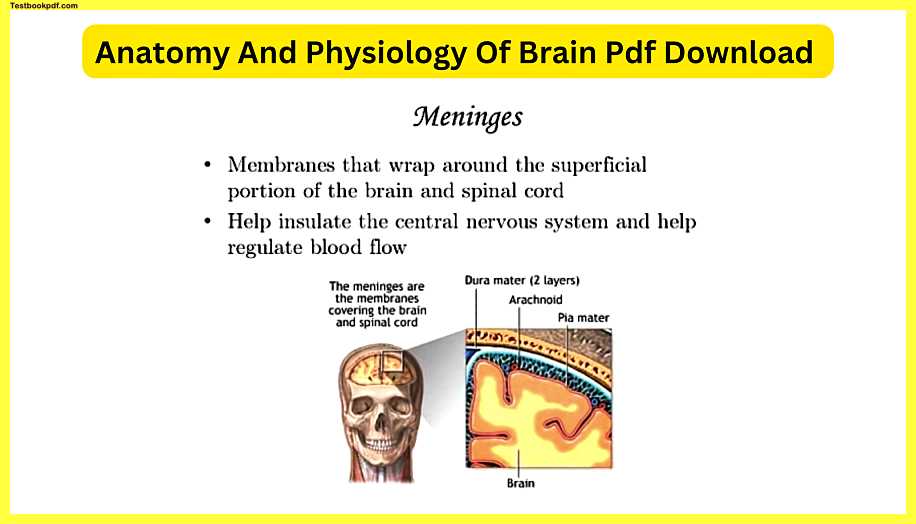
The meninges are wrappings around the brain and spinal cord so it really envelops wrap around the central nervous system as a whole and it’s around the superficial portion of the outside it helps insulate the central nervous system and helps regulate blood flow sometimes you get what’s called meningitis which I’m gonna mention again in a little bit that’s an infection of these meninges and the problem with that is if we zoom in to where the meninges are here is a part of what looks to be either the parietal bone or frontal bone and just deep to it you have these membranes that are right in between the skull and the brain if you get bacterial virus fungus in this part of your head your immune response to that being there is going to create inflammation swelling and the problem with swelling here is that your skull is not gonna budge but the swelling in this region is going to start pushing on your cerebral cortex and that’s going to be potentially fatal.
So meningitis is one of those things that you gotta get treated as soon as possible but besides the infections, the meninges are very important by the way the singular version of this word it’s not often used but they say many Mining but I hear the word meninges a lot more because we’re talking about them as a whole there are several layers the dura mater is the one that’s just adjacent to the inside of the skull so the right under the cranium you’ve got this area called the dura mater and then you have another layer that’s just deep to that and there’s a little pocket it in between the two layers of the dura mater that’s where the CSF is gonna be.
So the flowing of that cerebrospinal fluid around the brain and spinal cord is gonna be within that dura mater and then you have this area called the arachnoid between the dura mater and then the Pia mater is right on top of the cerebral cortex that’s the part of amenities that is directly adjacent to that gray matter of the cerebrum and the way that I keep them straight is I think of it once again like I’ve said before in alphabetical order dura mater D comes before P so I think from top to bottom most superior or most superficial to deep and so it’s the two layers of dura mater and then the Pia a way to remember that dura mater has two layers is d for dye meaning 2 D for dura mater meaning two whatever works so these are the layers of the meninges that are protecting the such a nervous system and helping regulate blood flow.
Brain disorders and conditions
Finally some brain disorders and conditions like I mentioned previously seizures most common in people with epilepsy but a seizure can happen in anyone not all is known about the origination of seizures but there’s a lot of studies being done it’s kind of like an electrical storm in the brain and the reason why you know the someone with the seizure usually ends up on the ground convulsing is because it’s stimulating so many parts of the brain that are typically regulating motor control that it’s out of control you don’t have that coordinated inhibition and excitation of those muscles so it ends up you know creating a situation where they’re convulsing a piece of advice do not try to prevent someone from swallowing their tongue if they’re having a seizure when a seizure is happening because there’s an uncoordinated movement sometimes the tongue can get in the way of the respiratory passageways but that’s not as common as you would think people who’ve tried to prevent someone from swallowing their tongue while they’re seizing have getting their faith they’ve gotten their fingers bitten off you know and that’s that’s not something you want to do you don’t to put your fingers in a person’s mouth who can’t control consciously their skeletal muscle movements cuz they might be biting down what you should do is find a way to cradle that person’s head get a pillow get something hot soft or just hold their head to prevent their head from hitting something hard and when the seizure is done assess how they’re doing
Concussions
Concussions are fairly common, especially in sports that involve collisions car accidents are another way that some can get a concussion but anytime your head hits something very hard or something hits your head very hard it can cause a brain bruise that’s basically what a concussion has to do with your head being hit so hard that all of them.
Meningitis
The Meningitis cushioning your brain the cerebral spinal fluid being that nice buffer between your brain and the inside of your cranial cavity wasn’t enough your brain physically hit into your skull and usually it’s just damage on the superficial parts of the brain that someone does recover from now enough concussions over time it can cause more permanent damage meningitis already mentioned before either bacterial viruses or funguses fungi I should say I can actually infect your Meningitis the signs of meningitis happening is fever you know a sense of pain up here it’s like a really bad headache confusion kind of not a sense of what’s going on and the other one is if the person has trouble doing this if they get a lot of pain from moving their head down to their chest it’s because they’re stretching part of them and in geez and that activity will cause a lot of pain if those signs are happening to go to the emergency room better to be safe.
Stroke
Than stroke also knows a cerebrovascular accident has to do when arteries going up into the brain get clogged depending on what artery gets clogged or arterioles smaller arteries that’s going to cut off blood flow to a certain part of the brain strokes can be fatal sometimes they’re not it really just depends a sign that someone has had a stroke is you get kind of a disagreement of what the sides of the body are doing or what they’re able to do because a stroke is gonna happen on the right or left side it’s very rare that you’d have two strokes and then left the right side happening simultaneously
So let’s say a person has a stroke in their left hemisphere that’s gonna affect the right side of their body so if you ask the person to smile and they only seem to be able to like to lift up this part of the mouth that’s a sign they’ve had a stroke or if you ask the person to lift up both of their arms if they just can’t lift it quite as well that’s another sign that they’ve had a stroke and that you should take them to the hospital
Hemorrhage
this is not just in the brain but you can get hemorrhages in the brain it’s basically internal bleeding you know you can have hemorrhages on the surface of the brain have hemorrhages within the brain depending on how bad they are they can be fatal and really surgery is required to get rid of that bleeding.
Aphasia
aphasia is anytime that a person has the inability to speak or read at a normal level and that has to do with underdevelopment of those regions or damage to those regions because of an accident related to that is dyslexia.
Dyslexia
I’ve heard estimates that it affects as much as fifteen percent of children and it still affects adults too dyslexia is not something that’s incurable it’s something that someone can get over but it has to do with reading and the words not completely making sense as it does in a normal reader so let’s say the person is looking at the word pie they may actually see e IP when they read it and that’s, of course, gonna create some issues with the understanding there are therapies that the person can do to get to the point where they see this as they should so it’s something that with work you can conquer it.
Disconnection syndrome
Disconnection syndrome is a result of like I mentioned earlier cutting through the corpus callosum having the left and right hemispheres not be in communication and having sort of that disagreement of what the left and right hands or left the right sides of the body are telling a person like I also mentioned earlier there is a small area anteriorly connecting parts of the frontal lobes that can still be in communication but the majority of that communication is definitely in the corpus callosum.
Hydrocephalus
Hydrocephalus literally means water on the brain and this is most common in infants if they are not regulating the amount of CSF cerebral spinal fluid within the brain and on the outside of the brain and spinal cord if they’re producing way too much of it you can get this swelling almost looking like their head is ballooning and that’s most common in an infant because if you saw the skeletal system lessons those cranial bones are not completely fused yet so there’s a little bit of wiggle room and that buildup of CSF looks like the head is ballooning and it can be fatal if it’s not treated
Parkinson disease
I mentioned earlier has to do with uncoordinated motor stimulation and has to do a little bit at least a little bit with dopamine imbalance there isn’t a complete cure yet I believe one day there will be a solid cure for Parkinson’s because it’s probably more than just dopamine there are possibly genetic factors and environmental factors that have to do with Parkinson’s disease and it’s onset.
Alzheimer’s disease
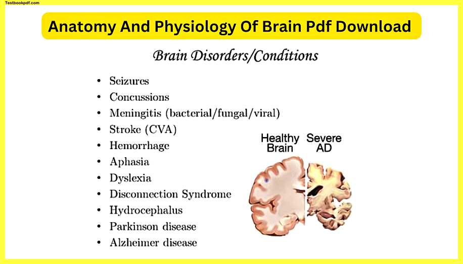
I’ve heard people mispronounce as they say old-timers it’s Alzheimer’s and it’s named after a doctor that researched it for many years Alzheimer’s disease here is a picture that shows you what it looks like here’s a healthy brain one of those coronal or frontal sections through the cerebrum and this is severe AD or Alzheimer’s disease and you can see that there’s a lot of deterioration it’s going to affect the hippocampus and that’s why with severe AD is gonna have memory problems or a sense that maybe they’re in a previous time one of my great-grandparents who had Alzheimer’s when he would talk to her she would think a lot about previous times as if she was still in the moment and she would look at relatives that she knew and say oh you remind me of this person as if she was stuck in a previous time and then of course the cerebral cortex you could see the outsides of the cerebrum are severely impacted you’d see kind of a wearing away of a lot of that gray matter and that’s gonna gradually incapacitate somebody to the point where they’re not gonna be able to take care of themselves there’s a lot of research into Alzheimer’s disease identifying factors that make it more likely for someone to get it and once again genetics has something to do with it but there may be certain environmental factors that make it more likely for one person or another to get it and of course as you age the chances of ad coming into play increase so if you look at a population of people who are in their 60s they’re less likely to get it some people here 60s can get it but when you look at a population their 80s or 90s the incidence of Alzheimer’s goes up significantly.
Thanks for reading this Complete article.
